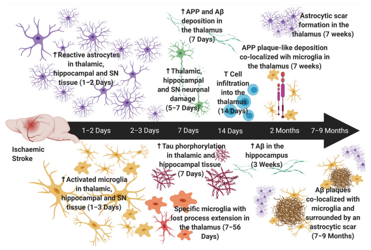
Figure 1.
Neuroinflammation in Secondary Neurodegeneration following Experimental Stroke. Astrocytes are first increased within the thalamus, hippocampus and substantia nigra (SN) at ~one to two days post-stroke [32,185,186]. Astrocytic scar formation is first apparent in thalamic nuclei at seven weeks post-stroke [51]. Activated microglia are first increased within the thalamus, hippocampus and SN at one to three days post-stroke [177], with specific microglia (loss of process extension with intact phagocytotic functioning) seen in the thalamus at seven days and up to fifty-six days post-stroke [173,202]. Neuronal damage is noted after glial reactivity (~five to seven days) [185,186,199]. Tau phosphorylation in the thalamus [46] and hippocampus [48], and Aβ and APP deposition in the thalamus, is observed at seven days post-stroke [43]. Conversely, Aβ in the hippocampus is not apparent until three weeks post-stroke [47]. T cell infiltration into the thalamus is observed at fourteen days post-stroke [184]. APP deposition adopted plaque-like morphology and was colocalized with microglia at seven weeks post-stroke and Aβ plaques were colocalized with microglia at seven months [174] and was surrounded by an astrocytic scar at nine months post-stroke [43]. ‘↑’ denotes an increase. Created with BioRender© (https://biorender.com) (accessed on 30 November 2021).

