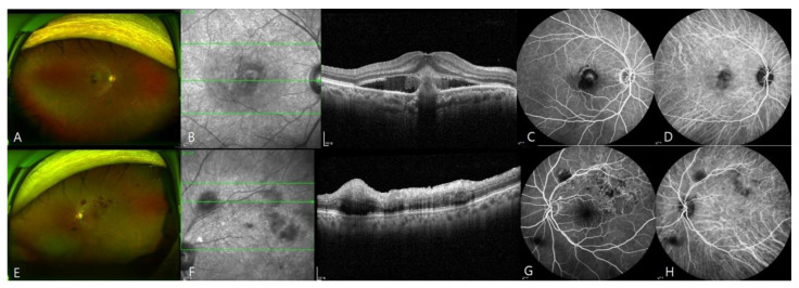
Figure 1.
Images of a patient with submacular hemorrhage and retinal vein occlusion (RVO) in both eyes (eyes 7 and 19). The patient developed submacular hemorrhage in the right eye and RVO in the left eye at 2 weeks after the first injection of BNT162b2. (A–D), Wide-field fundus photograph, OCT scan, fluorescein angiograph, and ICG angiograph of the right eye. OCT revealed subretinal hemorrhage. (E–H), Ultra-wide-field fundus photograph, OCT scan, fluorescein angiograph, and ICG angiograph of the left eye. Fluorescence angiography revealed blocked fluorescence at the site of retinal hemorrhage.

