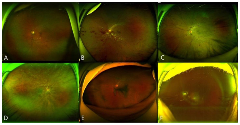
Figure 3.
Images of representative eyes exhibiting retinal vein occlusion (RVO) after vaccination. (A,B), Wide-field fundus photographs revealing branch RVO (eyes 14 and 21). (C,D), Wide-field fundus photographs revealing central RVO (eyes 23 and 17). (E,F), Wide-field fundus photographs of RVO with vitreous hemorrhage (eyes 22 and 13).

