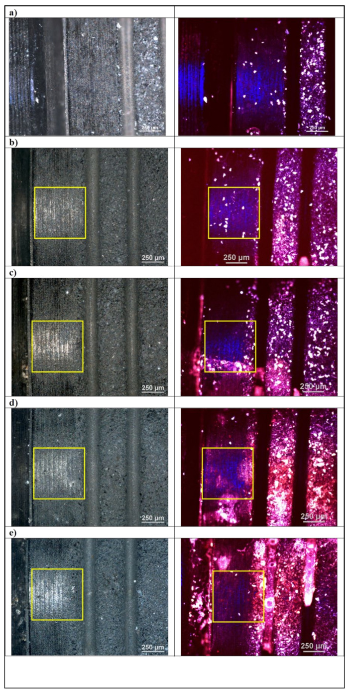
Figure 1.
Optical and confocal microscopy. Micrograph of the implant sample surface (left column) under optical microscope and (right column) under confocal microscope, laser wavelength λ = 405 nm. (a) randomly selected site, (b) control, (c–e) disinfection variants. Laser application sites are marked with a frame.

