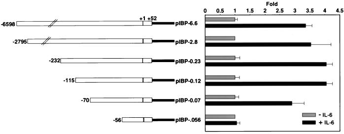
FIG. 2.
Functional analyses of the mouse IGFBP-1 promoter in HepG2 cells and deletion mapping of the mouse IGFBP-1 promoter. Left, schematic diagrams of the various mouse IGFBP-1 deletion constructs; right, graphical representation of relative luciferase activity after normalization to β-galactosidase activity. To determine enzyme activity, 0.5 μg of the indicated reporter and 1 μg of pRSV-β-galactosidase were transfected in HepG2 cells by the calcium phosphate precipitation method using the 60-mm-diameter dishes. The cells were treated with rhIL-6 (100 ng/ml) for 4 h. The luciferase activity was expressed as fold induction relative to the basal activity of the reporter construct in the absence of IL-6 treatment. Six independent determinants were made for each construct by performing duplicates in three separate experiments. The values were plotted as averages ± standard deviations.

