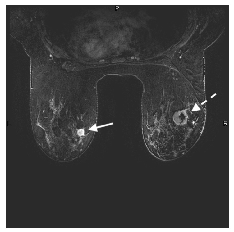
Figure 1.
28-year-old woman with a known BRCA 2 genetic mutation presented for her baseline annual high-risk screening MRI. Axial contrast-enhanced T1-weighted fat-suppressed image shows a 1.4 × 1.3 × 1.2 cm rim enhancing mass in the upper inner left breast (solid white arrow). This was a biopsy-proven grade 3 invasive ductal carcinoma. There is also a 3.1 × 3.6 × 2.1 cm rim enhancing mass with central necrosis in the upper outer right breast (white dashed arrow). This was also biopsy-proven grade 3 invasive ductal carcinoma.

