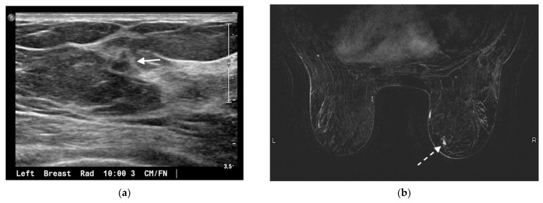
Figure 2.
75-year-old woman presented for annual screening evaluation. Mammogram demonstrated a left breast asymmetry (not shown) which prompted further workup. (a) Left breast ultrasound in radial projection at the 10 o’clock axis 3 cm from the nipple demonstrates a suspicious 0.5 × 0.6 × 0.4 cm hypoechoic antiparallel mass with irregular margins and posterior acoustic shadowing (arrow), corresponding to the mammographic abnormality. Ultrasound guided biopsy yielded invasive ductal carcinoma, mucinous type. (b) MRI was performed to evaluate the extent of disease; axial contrast-enhanced T1-weighted fat-suppressed image demonstrates a separate 0.7 × 0.5 cm enhancing irregular mass in the lower inner quadrant of the contralateral right breast (dashed arrow). This was biopsied under MRI guidance and yielded invasive ductal carcinoma, mucinous type. The known left breast cancer is not shown.

