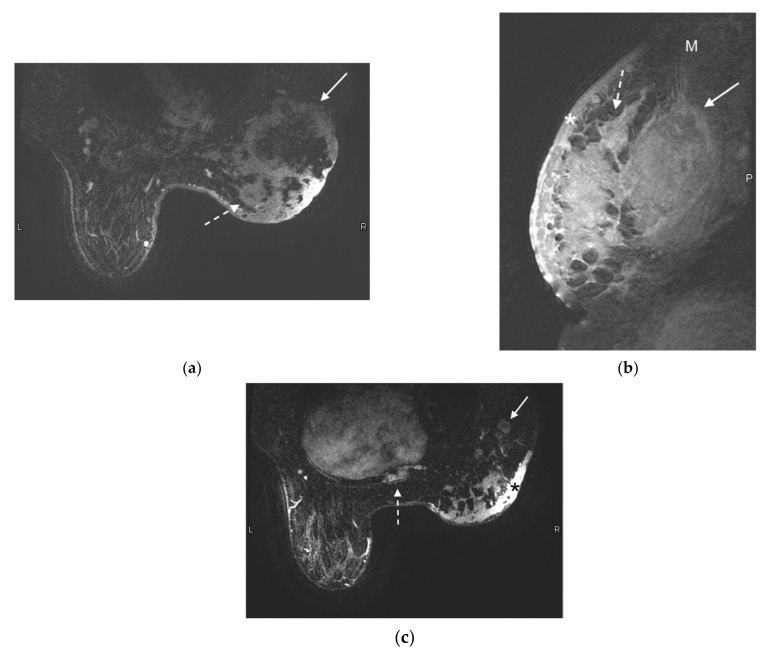
Figure 3.
55-year-old woman presented with a palpable breast mass and asymmetric breast size. Mammogram and ultrasound showing a large mass are not shown. MRI was performed to evaluate for extent of disease. (a) Axial contrast-enhanced T1-weighted fat-suppressed image demonstrates a 13 cm ring-enhancing centrally necrotic lateral right breast mass (solid arrow). Additional rim-enhancing centrally necrotic satellite lesions are noted at the medial aspect of the breast (dashed arrow). (b) Sagittal contrast-enhanced T1-weighted fat-saturated image demonstrates a large irregular heterogeneously enhancing right breast mass invading the right pectoralis muscle (arrow). The edge of the normal pectoralis muscle (M) is seen superiorly. Diffuse skin thickening and enhancement (*) is compatible with skin involvement, and satellite lesions (dashed arrow) are redemonstrated. p demarcates the posterior aspect of the image. (c) Axial contrast-enhanced T1-weighted fat-saturated image demonstrates abnormally enlarged and enhancing right axillary lymph nodes (arrow) and an enhancing metastatic bone lesion in the sternum (dashed arrow). Right breast skin thickening (*) is again noted.

