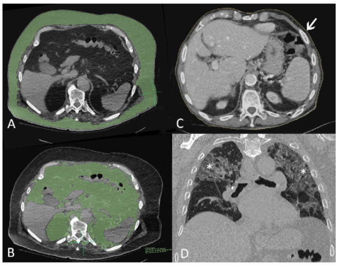
Figure 1.
Computed tomography (CT) images of three patients with COVID-19 pneumonia. (A,B), axial images of 89-year-old man, soft tissue window setting. The first slice caudal to the pleural recesses shows overlay segmented parietal (A) and visceral (B) fat in green. (C) axial image of 93-year-old man, soft tissue window setting. Fine yellow line (arrow) delineates body circumference on the first slice caudal to pleural recesses. (D) Coronal-oblique reconstructed image of 86-year-old man in lung window setting shows stage 3 lung infiltrates with ground glass opacities, intralobular septal thickening, and parenchymal bands (asterisks).

