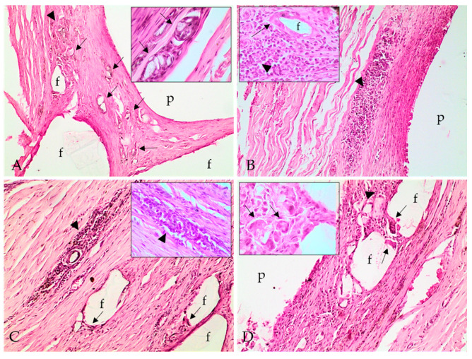
Figure 4.
Photomicrographs of implant polymer sites with the polymer (p), lymphoplasmacytic inflammatory infiltrate (arrowhead), and epithelioid macrophages and multinucleated giant cells, with intracytoplasmic polymer material, thus characterizing polymer phagocytosis (arrows). (A) Inflammatory infiltrate and polymer phagocytosis at 24 weeks; (B) 28 weeks; (C) 34 weeks; and (D) 38 weeks. Hematoxylin and eosin stain; 100×. Inlet (A,D): multinucleated giant cells with intracytoplasmic fragments of polymer material, characterizing phagocytosis (arrows). Inlet (B,C): lymphoplasmacytic inflammatory infiltrate (arrowhead). Inlet (C): epithelioid macrophages (arrow) delimiting a polymer fragment (f). Hematoxylin and eosin stain; 400×.

