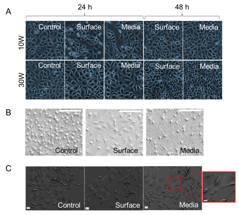
Figure 5.
(A) Phase contrast images of wells containing fibroblasts grown in the presence of the coatings after 24 h and 48 h of incubation, scale bar 100 µm, and (B) SEM images of fibroblasts attached on the surfaces of substrata after 48 h of incubation, scale bar 50 µm. (C) SEM images of osteoblasts attached on the surfaces of substrata after 48 h of incubation, scale bar 20 µm, scale bar for inset 10 µm. For all images: “Control” denotes as-deposited surface and standard media, “Surface” is plasma treated surface and standard media, “Media” is as-deposited sample and plasma treated media. Data shown for polymer coatings fabricated at 10 W.

