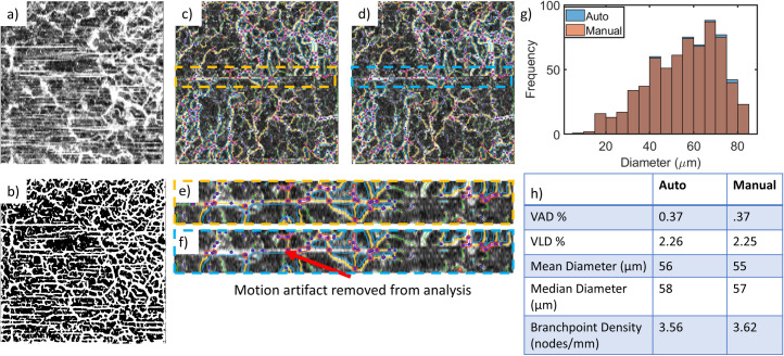
Fig 8. Motion artifacts, poor-quality images, and manual curation.
Images with significant motion artifact (a) must be removed from the analysis since the segmentation algorithm cannot distinguish between the vessels and motion artifacts; the resultant segmented OCTA MIP (b) does not accurately represent the microvascular architecture. For images with minimal motion artifacts, such as the images in (c) and (d), the individual motion artifacts can be removed manually. (e) and (f) show insets of (c) and (d), respectively, demonstrating the removal of the motion artifact using manual curation. Depending on the VAD, individual motion artifacts likely have little impact on the generated metrics, as shown in the histogram of diameters (g) and the table of generated metrics with and without manual curation (h). Note that in (g), the columns representing the results with manual curation are always equal in height or shorter than the automatically generated values since no vessels were added by manual curation in this instance. Square OCTA MIP images in (a-d) are 5 mm × 5 mm and comprise an axial range of 500 μm. The images in (e) and (f) are 0.65 mm × 5 mm.

