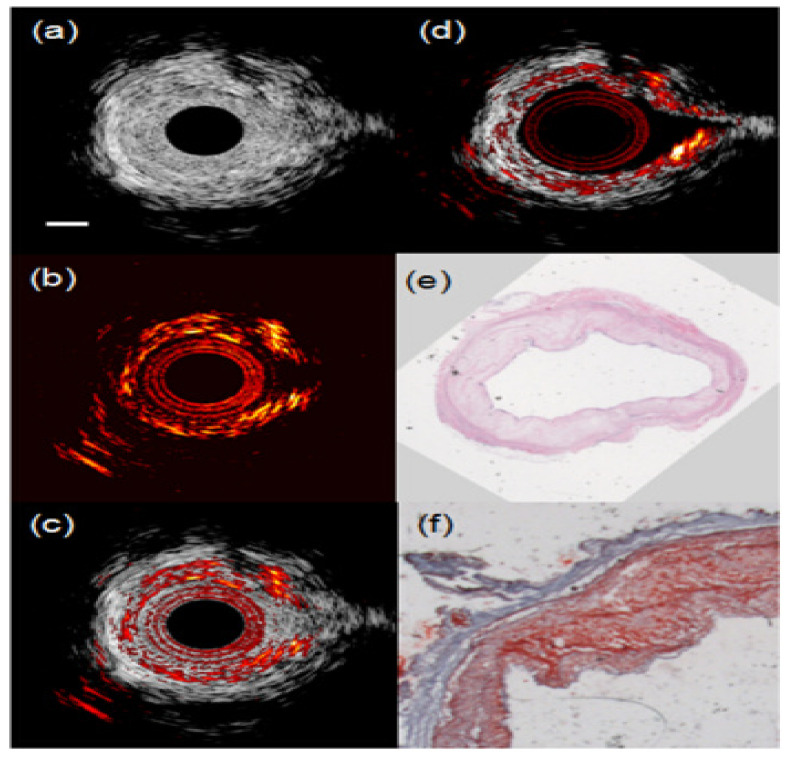
Figure 6.
(a) IVUS, (b) IVPA, and (c) combined IVUS/IVPA images of an atherosclerotic rabbit aorta acquired in the presence of blood. (d) Combined IVUS/IVPA image of the same cross section of the aorta imaged in saline. IVUS and IVPA images are displayed at 35 dB and 20 dB, respectively. The scale bar is 1 mm. (e) H&E and (f) Oil red O stain of the tissue slice adjacent to the imaged tissue cross section indicate that the aorta has lipid-rich plaque. (Reprint from [122] with permission).

