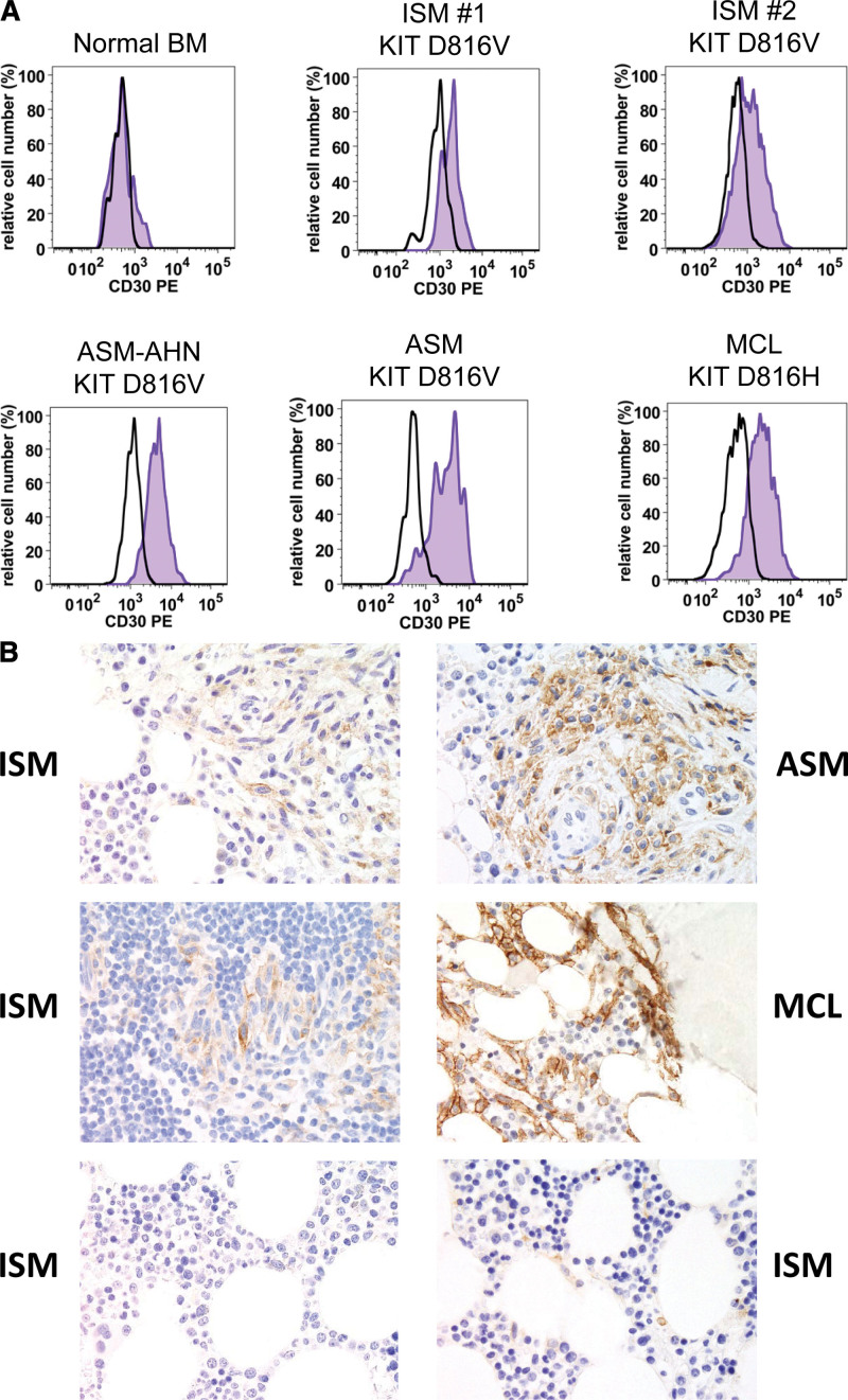
Figure 1.
Expression of CD30 in neoplastic mast cells in systemic mastocytosis. (A), Flow cytometric detection of CD30 on neoplastic MC in patients with SM. BM cells were obtained from a control patient (no known BM disease; upper left image), patients with indolent SM (ISM: upper middle and right panels), and patients with advanced SM, namely 1 with ASM with an AHN (ASM-AHN: lower left image), 1 with ASM (lower middle histogram), and 1 with MCL by multicolor flow cytometry on a FACSCanto (Becton Dickinson). MC were identified as CD117++/CD45+/CD34− cells and stained with a phycoerythrin-labeled monoclonal antibody against CD30 (BerH8 from BD Biosciences; blue histograms). The isotype-matched control antibody (black open histogram) is also shown. (B), Immunohistochemical detection of CD30 in neoplastic MC. BM biopsy sections from patients with ISM, ASM, and MCL (as indicated) were stained with a monoclonal antibody against CD30 (Ber-H2 from Dako, Glostrup, Denmark) by an indirect immunoperoxidase staining technique as reported.81 The 2 images at the bottom show the nonaffected BM in 2 patients with ISM (control). Images were prepared using an Olympus DP21 camera connected to an Olympus BX50 microscope equipped with 60×/0.90 UPlan-Apo objective lens (Olympus, Hamburg, Germany) and processed with Adobe Photoshop CS2 software version 9.0 (Adobe Systems, San Jose, CA) as described.81 All patients gave written informed consent before BM samples were obtained and analyzed. AHN = associated hematologic neoplasm; ASM = aggressive SM; BM = bone marrow; ISM = MC = mast cells; MCL = MC leukemia; SM = systemic mastocytosis.

