Figure 7.

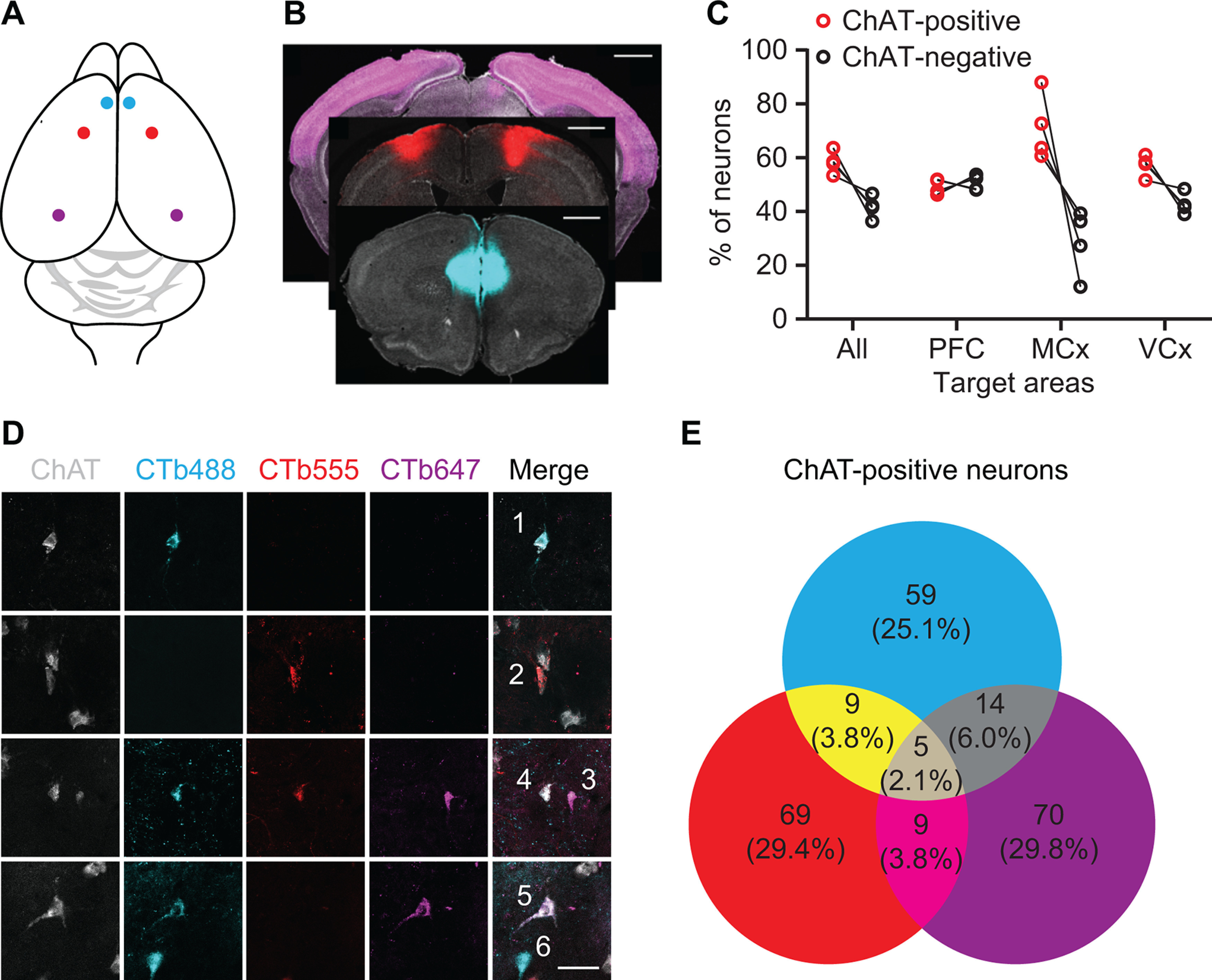
Retrograde labeling of NBM/SI cholinergic projections to cortex. A, Illustration of retrograde tracing strategy: CTB conjugated to Alexa Fluor 488, 555, or 647 was bilaterally injected to mPFC (cyan), motor cortex (red), or visual cortex (purple), respectively. B, CTB injection sites: mPFC (cyan), motor cortex (red), and visual cortex (purple). Sections were stained with DAPI (gray; scale bar: 1 mm). C, Quantification of cholinergic and noncholinergic neurons projecting to cerebral cortices. D, Retrograde labeling of NBM/SI with CTB conjugates. Neurons 1, 2, and 3 projected to one of these three cortices. Neuron 4 projected to mPFC and motor cortex. Neuron 5 projected to mPFC and visual cortex. Neuron 6 is noncholinergic (scale bar: 40 μm). E, Quantification of total CTB-labeled cholinergic NBM/SI neurons.
