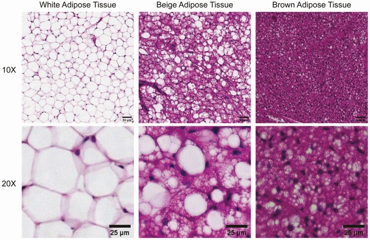
Figure 2.
Morphological features of brown, beige, and white adipocytes in adipose tissues. White adipocytes in white adipose tissue, exemplified by mouse gonadal fat (left) are larger and contain a single lipid droplet per cell. Brown adipocytes in mouse interscapular brown adipose tissue (right), are smaller in size and contain numerous lipid droplets. Beige adipocytes in inguinal fat of cold-exposed mice (middle), are intermediate in size and have characteristics of both white and brown adipocytes, having a large droplet and multiple small droplets in the same cell, or multiple small droplets. Bottom row displays a higher magnification image of each tissue type. Scale bar: 50 μm (top row); 25 μm (bottom row).

