FIGURE 3.

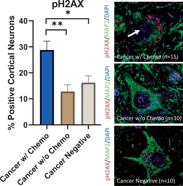
Elevated DNA damage in cortical neurons after chemotherapy. (Left) An increased percentage of frontal lobe cortical neurons in cancer patients treated with chemotherapy contain nuclear pH2AX-positive foci (28.8%) compared to frontal lobe cortical neurons in cancer patients not treated with chemotherapy (12.8%, p < 0.01) and patients without history of cancer (16.2%, p < 0.05). Error bars represent the standard error of the mean. (Right) Representative pH2AX and MAP2 double-labeled immunofluorescent images from all 3 cohorts counterstained with DAPI; the top panel shows a neuron with a pH2AX-positive focus (arrow) in a cancer patient treated with chemotherapy; the middle and bottom panels show neurons without pH2AX-positive foci in control patients. Nonspecific pH2AX immunopositivity is seen in cytoplasmic lipofuscin granules. DAPI, 4',6-diamidino-2-phenylindole; MAP2, microtubule-associated protein 2; pH2AX, phospho-H2AX; *p < 0.05, **p < 0.01.
