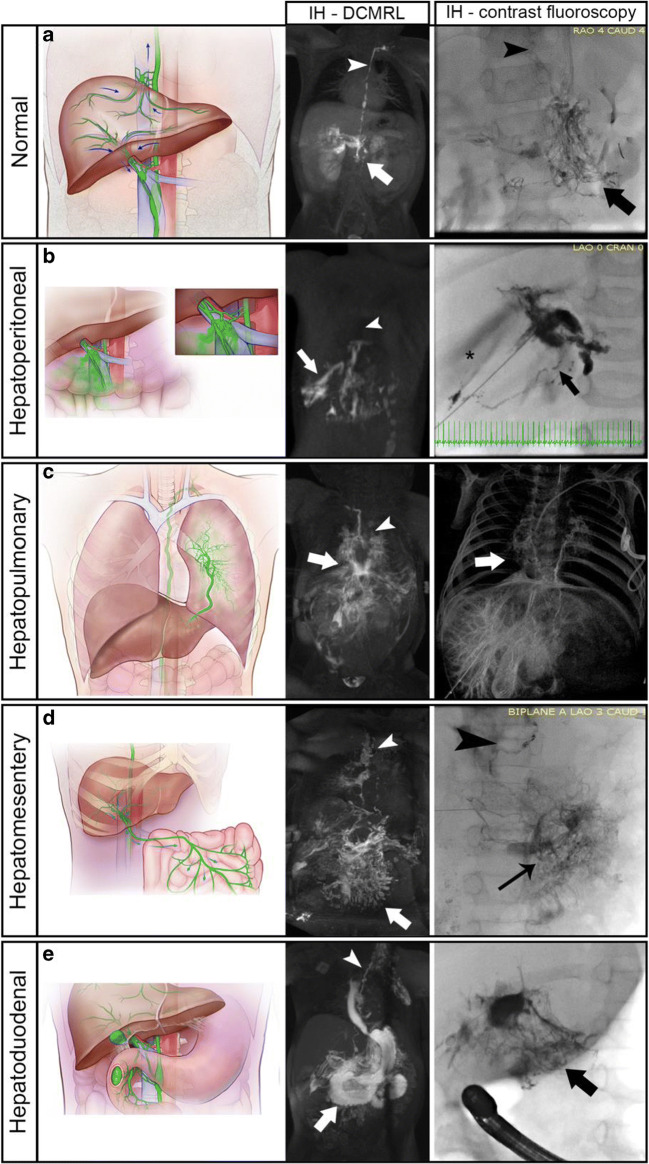
Fig. 1.
Normal and abnormal hepatic lymphatics with representative maximum intensity projections (MIP) of IH DCMRL and IH contrast fluoroscopy images. Arrowhead represents the normal thoracic duct and arrows denote the abnormal lymphatic connections. (a) Normal lymphatic drainage diagram of superficial and deep liver lymphatic drainage. Superficial (capsular) lymphatics directly enter the central TD near the diaphragm while deep (peri-portal) lymphatics course toward the liver hilum and toward the celiac and pancreatic lymphatic networks (arrow) with further connections to the cisterna chylii and thoracic duct (arrowhead). (b) Hepatoperitoneal connections with disruption of liver lymphatics after exiting the liver hilum. (c). Hepatopulmonary connections from the pericapsular lymphatics of the left liver lobe to the left mainstem bronchus. Arrows represent the abnormal connections with the ductal remnant noted with an arrowhead (d) Hepatomesenteric connections from the liver to the mesentery with intact TD and pulmonary lymphatic perfusion (e) Hepatoduodenal representation of the liver lymphatics as they exit the liver hilum and course to the inner curvature of the 1st – 3rd portions of the duodenum (arrows) with significant reflux into the stomach and esophagus (asterisk) with propagation forward to the proximal jejunum

