Fig. 3.
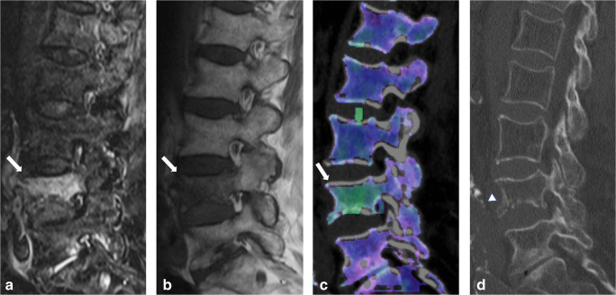
Eighty-seven-year-old woman presenting with acute spinal trauma due to a domestic fall. a Sagittal turbo inversion recovery magnitude (TIRM)–magnetic resonance imaging (MRI) series, b spin-echo (SE) T1-weighted MRI series, and c dual-energy computed tomography (DECT)–virtual non-calcium (VNCa) reconstructions showing bone marrow edema (BME) in all four quadrants of L4 (arrow). All readers were concordant in assessing BME presence (score 3 = distinct BME) and extent (score 4 = all quadrants) on both techniques. d In addition, sagittal conventional grayscale DECT images allowed for detection of an acute slightly dislocated fracture of the ventral ground plate of L1 (arrowhead) in terms of a tear drop fracture with potentially associated instability by all readers in this study. Confidence in depicting fracture lines was rated as intermediate (score 2) and high (score 3) by 5/5 readers on MR and DECT image series, respectively

