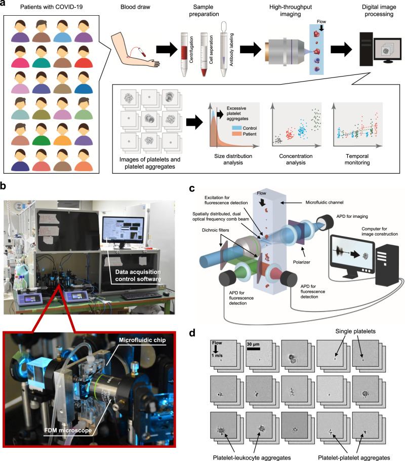
Fig. 1. Massive image-based profiling of circulating platelets and platelet aggregates at single-cell resolution.
a Experimental workflow consisting of blood draw, sample preparation, high-throughput imaging, and digital image analysis. b Pictures of the FDM microscope on the microfluidic chip installed in the Department of Clinical Laboratory at the University of Tokyo Hospital. The inset shows an enlarged view of the microfluidic chip with the FDM microscope. c Schematic of the FDM microscope for high-throughput, blur-free, bright-field image acquisition. APD: avalanche photodetector. d Typical bright-field images of single platelets and platelet aggregates acquired by the FDM microscope. For information about data reproducibility, see “human subjects” and “statistical analysis” in the Methods section for details.

