FIGURE 2.
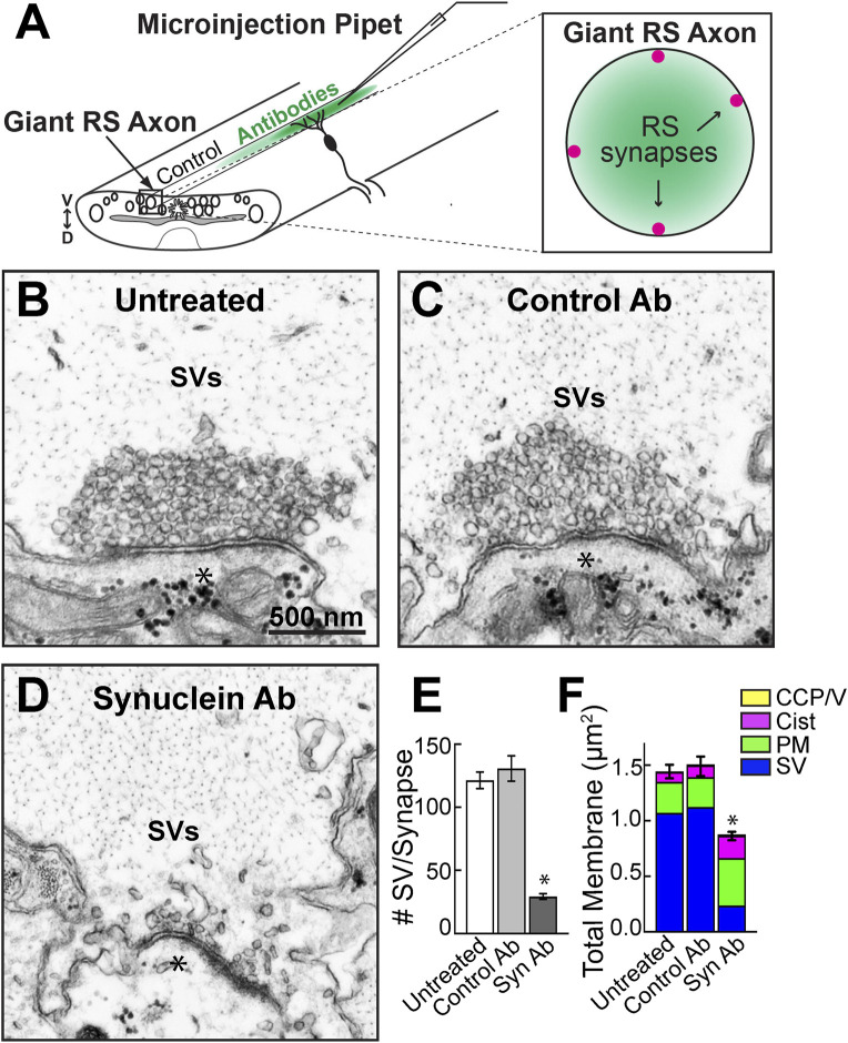
Microinjection of a pan-synuclein antibody induced a severe depletion of synaptic vesicles at resting lamprey synapses. (A) Diagram showing acute perturbation strategy. Antibodies are injected directly into the giant RS axons (left), which delivers the reagents directly to the synaptic vesicle clusters at resting RS synapses (right). Note the concentration gradient of injected antibodies, which permits evaluation of dose-dependent effects. (B–D) Electron micrographs of giant RS synapses. At untreated synapses, or after injection with Control IgG antibodies (Ab), the synaptic vesicle (SV) clusters are large and tightly clustered. After treatment with the Synuclein Ab, the SV clusters were severely depleted. Asterisks mark postsynaptic dendrites. Scale bar in B is 500 nm and applies to C–D. (E) Compared to controls, the synuclein antibody reduced the number of SVs in the vesicle cluster by >75%. Data are plotted as mean/section/synapse. (F) The synuclein antibody also significantly reduced the total amount of membrane at synapses, primarily due to the loss of membrane associated with SVs. Moderate increases in plasma membrane (PM) and cisternae (cist; putative endosomes) were observed. Bars in E and F represent mean ± SEM from n = 23–62 synapses, 2-5 axons/animals. Asterisks indicate statistical significance (p < 0.0001) by ANOVA, Tukey post hoc.

