FIGURE 3.
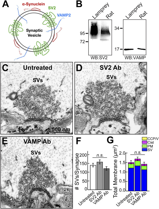
Microinjection of SV2 and VAMP antibodies had no noticeable effect on synaptic vesicle clusters. (A) Diagram of a synaptic vesicle showing α-synuclein and two other vesicle-associated proteins, SV2 and VAMP. (B) Western blots against SV2 and VAMP show bands of the expected molecular weights in both lamprey CNS and rat brain protein lysates. The SV2 band appears as a smear due to extensive glycosylation of the protein. (C–E) Compared to untreated synapses, no apparent changes in the morphologies of synaptic vesicle clusters were observed after treatment with either the SV2 or VAMP antibody. Asterisks mark the postsynaptic dendrites. Scale bar in C is 500 nm and applies to D-E. (F) SV2 and VAMP antibodies did not significantly impact the number of synaptic vesicles at synapses. Data are plotted as mean/section/synapse. (G) No changes were observed in the membrane distributions after treatment with the SV2 or VAMP antibody. Bars in F and G represent mean ± SEM from n = 24–38 synapses, 2-4 axons/animals. n. s. indicates “not significant” by ANOVA.

