Figure 2.
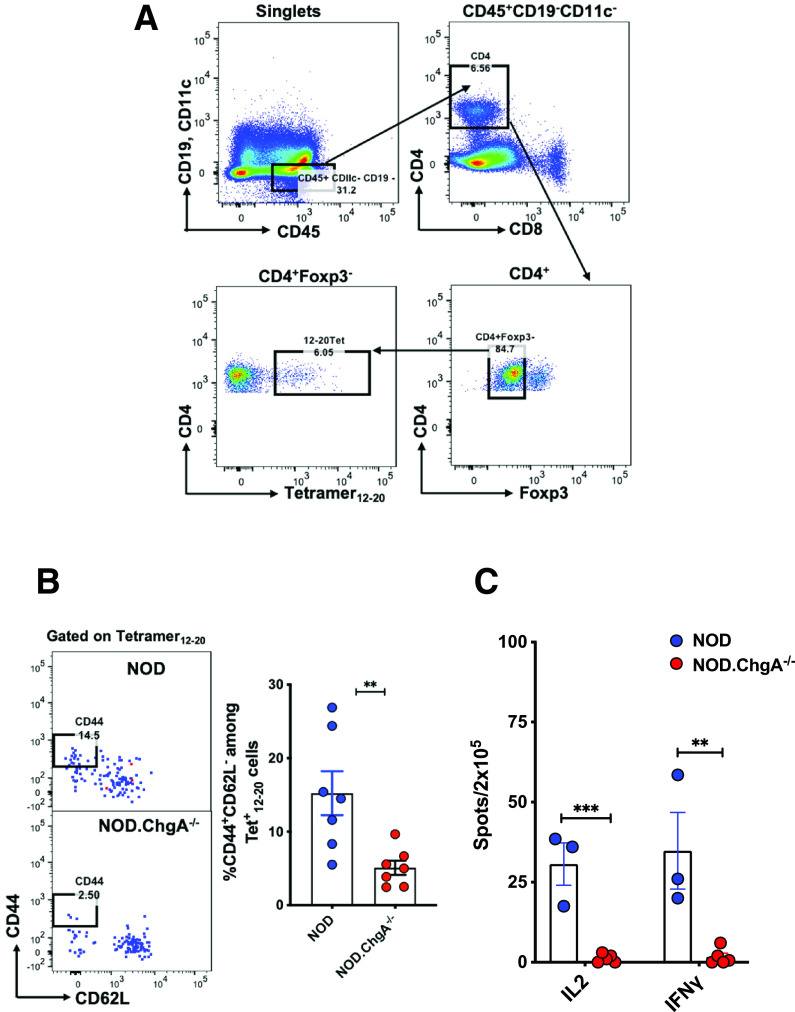
Defective priming of insulin-specific T cells in the secondary lymphoid tissues of NOD.ChgA−/− mice. A: FACS plots depicting the gating strategy of the InsB12–20 tetramer+ CD4 T cells in the secondary lymphoid tissue. B: Representative FACS plots showing the activation status of the InsB12–20 tetramer+ CD4 T cells in the secondary lymphoid tissues of NOD or NOD.ChgA−/− mice. Data show the percentage of the CD44+CD62L− cells among the InsB12–20 tetramer+ CD4 T cells as gated in panel A. The bar graph summarizes results from three independent experiments. Each dot represents individual mouse. C: ELISPOT assay showing IL-2 and interferon-γ (IFN-γ) production by CD4 T cells from the panLN cells of female NOD or NOD.ChgA−/− mice challenged and recalled with the InsB:9–23 peptide. Data (mean ± SEM) summarize results from three independent experiments. Each dot represents a biological replicate of panLN cells pooled from 3–5 mice. B and C: P values were calculated by unpaired two-tailed Student t test. **P < 0.01, ***P < 0.001.

