
FIGURE 3.
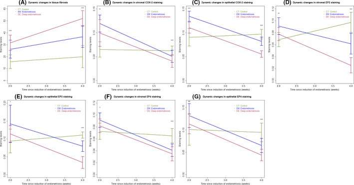
Summary result of immunohistochemistry and Masson trichrome staining in human samples showing the dynamic changes as endometriotic lesions progress over time. Dynamic changes in the extent of fibrosis in tissues/lesions in different groups of mice (A). Dynamic changes in the immunostaining of COX‐2 the stromal (B) and epithelial (C) components in tissues/lesions in different groups of mice. Dynamic changes in the immunostaining of EP2 the stromal (D) and epithelial (E) components in tissues/lesions in different groups of mice. Dynamic changes in the immunostaining of EP4 the stromal (F) and epithelial (G) components in tissues/lesions in different groups of mice. In all panels, the data are represented by the means ± SDs, and Kruskal's rank test was used. Symbols of statistical significance levels: *P < 0.05; **P < 0.01; ***P < 0.001; NS: not statistically significant (P > 0.05)