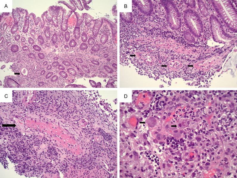
Figure 3.

A. From the first colonoscopy, sigmoid colon biopsy showed ischemic type changes with crypt atrophy (black arrow) and regenerative changes (10X objective). B. Three weeks later, cecal biopsy showed vasculitis (black arrow) with mildly active colitis in overlying colonic mucosa (20X objective). C. Ascending colon biopsy showed marked vascular thrombi (black arrow, 20X objective). D. Granulation tissue from transverse colon biopsy showed CMV viral inclusions (black arrow, 40X objective).
