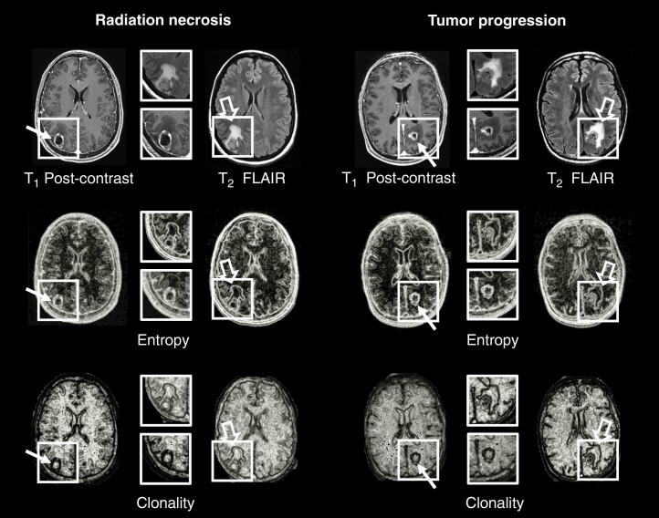
Figure 1.
Representative examples of radiation necrosis (Left) and tumor progression (Right) in metastatic brain lesions (boxes) treated with stereotactic radiosurgery. The original T1 post-contrast and T2-FLAIR images are shown in the top row. Radiomic images of entropy (middle row) and mpRad clonality (bottom row) demonstrate heterogeneity of the lesions (line arrows) and surrounding edema (open arrows).

