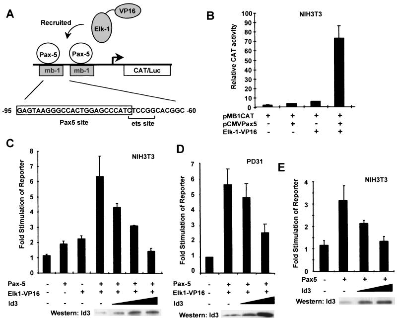
FIG. 7.
Id3 attenuates Pax-5-mediated activation of the mb-1 promoter. (A) Diagrammatic representation of the reporter system used, in which two copies of the indicated region from the mb-1 promoter drive the expression of a CAT or luciferase (Luc) gene. Pax-5 recruits ETS-domain proteins such as Elk-1 to this site (15). (B) Coexpression of Pax-5 and Elk-1–VP16 causes synergistic activation of the mb-1 promoter-regulated CAT reporter. NIH 3T3 cells were cotransfected with the mb-1–CAT reporter vector (1 μg) and with expression vectors encoding either Pax-5 (pAS1111; 0.25 μg) or Elk-1–VP16 (pAS348; 0.25 μg) alone or Pax-5 and Elk-1–VP16 together (0.25 μg each). (C) Id3 inhibits activation of the mb-1 reporter construct by Pax-5–Elk-1–VP16 complexes. NIH 3T3 cells were cotransfected with the mb-1–CAT reporter vector (1 μg), and expression vectors encoding either Pax-5 (pAS1111; 0.1 μg) or Elk-1–VP16 (pAS348; 0.1 μg) alone or Pax-5 and Elk-1–VP16 together (0.1 μg each). Where indicated, increasing amounts of the Id3 expression vector (pcDNA3Id3; 0.5, 2, and 4 μg) were cotransfected. (D) Id3 inhibits activation of the mb-1 reporter construct by Pax-5–Elk-1–VP16 complexes in B cells. PD31 cells were cotransfected with the mb-1–luc reporter vector and with expression vectors encoding Pax-5 (pAS1111; 5 μg) and Elk-1–VP16 (pAS348; 5 μg). Where indicated, increasing amounts of the Id3 expression vector (pcDNA3Id3; 5 and 20 μg) were cotransfected. (E) Id3 inhibits activation of the mb-1 reporter construct by Pax-5. NIH 3T3 cells were cotransfected with the mb-1–CAT reporter vector (1 μg), an expression vector encoding Pax-5 (pAS1111; 0.25 μg), and increasing amounts (0.5 and 4 μg) of an Id3 expression vector (pcDNA3Id3). Data are presented relative to those of the reporter plasmid alone (taken as 1). All values and standard errors were calculated from averages of duplicate (B, D, and E) or triplicate (C) samples and are representative of two or three independent experiments. In all cases, CAT-luciferase activity was measured 24 h after transfection. Gels (C to E) show Western blots of Id3 expression following transfection with increasing amounts of the Id3 expression vector.

