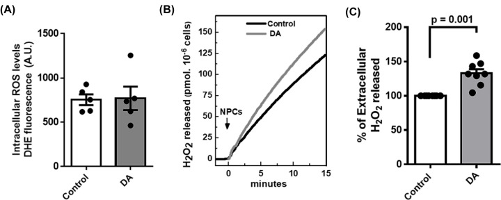
Figure 1. NPCs release higher levels of H2O2 upon dopamine treatment but no difference is detected in intracellular O●−.
(A) Quantification of the intracellular levels of O●- by DHE fluorescence of control and dopamine-treated NPCs (Dopamine 100 µM for 48 h). (B) Representative measurement of H2O2 release rate assessed fluorimetrically by Amplex Red-Peroxidase system, specific for H2O2, in intact NPCs. (C) Quantification of the rate of H2O2 release upon dopamine treatment when compared to control. In all experimental conditions we used 5 × 10⁴ cells/ml. The difference between groups was analyzed by unpaired t-test in a (n = 5 per group) and c (n = 7 per group). In c, we used Welches correction for unequal variances.

