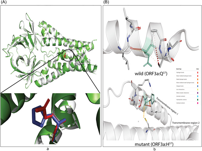
Figure 6.

The effect on transmembrane channel pore of ORF3a viroporin due to p.Q57H mutation. (A) The wild (Q57) and mutant (H57) ORF3a proteins are presented in light gray and green colors. The structural superposition displays no overall conformation change; however, the histidine at the position 57 of mutant ORF3a (deep blue) is slightly rotated from glutamine at the exact position of the wild protein (bright red). This change in rotamer state at the residue 57 may influence (B) the overall stability of H57 (upper part) overQ57 (lower part)because of ionic interaction of histidine (green; stick model) of transmembrane domain 1 (TM1) with cysteine at 81 (yellow stick) of TM2. The color code defined different bond types is shown in the inlet
