
FIGURE 2.
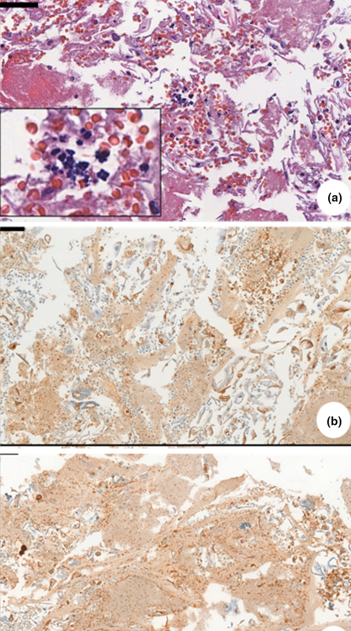
(a) Photomicrograph of a cell block of patient pictured in Figure 1h—routine hematoxylin and eosin (HE) stain. Sample of an ulcerative lesion in the lower lip. Sarcina ventriculi cocci characterized by multiple gram‐positive basophilic colored tetrad arrangement, scattered in epithelium. (b) Immunohistochemical of a cell block—anti‐HSV1 (abcam9533), Sample of an ulcerative lesion on dorsal tongue and buccal mucosa. Immunopositivity seen inside epithelial cell from basal layer