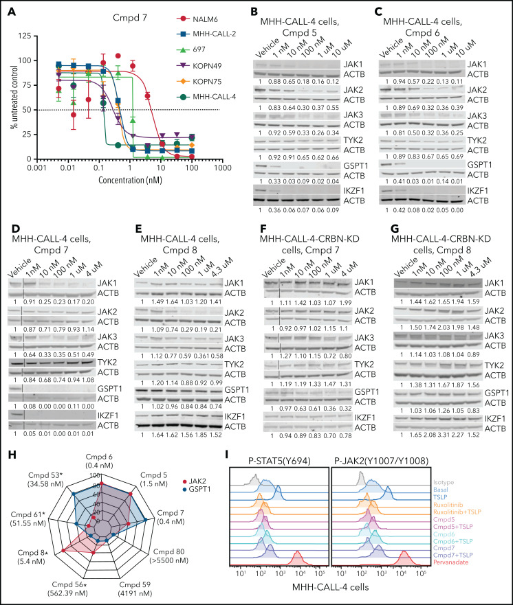
Figure 3.
Protein degradation, cytotoxicity, and effect of PROTAC compounds on JAK-STAT signaling pathway in MHH–CALL-4 cells. (A) Cytotoxicity of compound (Cmpd) 7 in 6 ALL cell lines (72-hours incubation). Dose-dependent degradation of JAKs, GSPT1, and IKZF1 by Cmpd 5 (B), Cmpd 6 (C), Cmpd 7 (D), and Cmpd 8 (E) in MHH–CALL-4 cells. Protein degradation by Cmpd 7 (F) and Cmpd 8 (G) in MHH–CALL-4 cells with CRBN knockdown (KD). (H) Spider chart showing 50% effective concentration of 9 representative PROTACs and degradation of JAK2 and GSPT1 in MHH–CALL-4 cells. Numbers within the web are the percentages of protein degradation normalized to vehicle control. The structures of Cmpd 53, 56, 59, 61, and 80 are shown in supplemental Figure 5. *50% Effective concentration was determined based on partial cytotoxicity curve; detailed information is summarized in supplemental Table 7. (I) Phosphoflow analysis of JAK-STAT5 signaling pathway in MHH–CALL-4 cells treated or not with 25 ng/mL TSLP. For inhibition, cells were treated with 1 µM ruxolitinib or PROTACs for 1 hour before TSLP stimulation.

