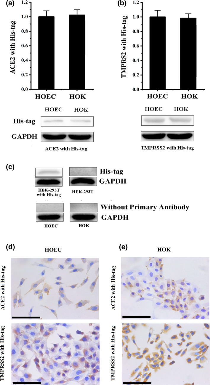
FIGURE 5.

The exogenous ACE2 and TMPRSS2 are absorbed to oral mucosa epithelial cells. (a) The results of the western blot confirmed that exogenous ACE2 was detected in HOEC and HOK cells. (b) The results of western blot confirmed that exogenous TMPRSS2 was detected in HOEC and HOK cells. (c) His‐tag was positive in HEK‐293T transfected with a His‐tagged Staphylococcus aureus cas9 and negative in HEK‐293T; HOEC and HOK, only stained by secondary antibodies, were negative. (d) The results of ICC confirmed that exogenous ACE2 was observed in the cytomembranes of HOEC and HOK cells. (e) The results of ICC confirmed that exogenous TMPRSS2 was observed in the cytomembranes of HOEC and HOK cells. (black scale bar = 100 μm) [Colour figure can be viewed at wileyonlinelibrary.com]
