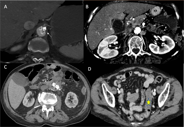
Fig. 1.

(a) Case 1: Aortic thrombosis at the origin of celiac trunk (arrow). (b) Case 1: Gangrenous gallbladder (arrow). (c) Case 2: Duodenum and abdominal aorta fused at the level of aneurism. Note fluid and air accumulation within aneurism cavity and aorto‐bi‐iliac endograft (arrow). (d) Case 3: Edema of the sigmoid colon (star) with perivisceral fluid accumulation
