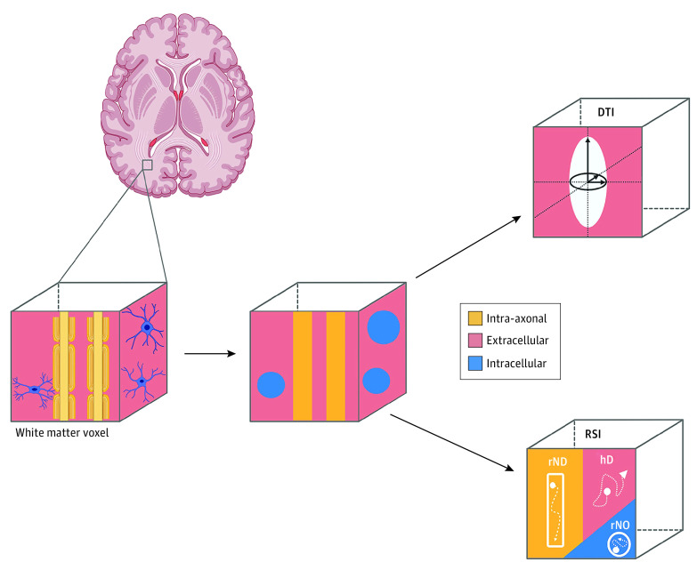
Figure 1. Diffusion-Weighted Imaging (DWI) Modeling Approaches.
Illustration of the biological components of white matter in an imaging voxel and schematic representation of the 2 diffusion tensor imaging (DTI) modeling approaches used in this study: DTI and restricted spectrum imaging (RSI). Diffusion tensor imaging measures extracellular water diffusion across a voxel. Primary DTI outcomes include fractional anisotropy (FA) and mean diffusivity (MD). Although the magnitude and direction of these outcomes allow for inferences to be made regarding axonal structure and integrity, DTI only allows for quantification of a single principal direction of diffusion and does not allow for characterization of the relative contribution of neurite orientations within a single voxel. In contrast, RSI is a biophysical model that allows for estimates of compartmentalized hindered and restricted water diffusion. Primary RSI outcomes include total hindered diffusion (hD) as well as restricted isotropic intracellular diffusion (rN0) and restricted directional intracellular diffusion (rND), which together provide greater insight into the biological properties of the microstructure of white matter tissue. The hindered compartment could encompass diffusion within intracellular spaces that allow for diffusion greater than the diffusion length scale (typically approximately 10 μm for human DTI). Given that rN0 and rND are normalized with respect to the hD compartments, changes in restricted compartments are relative to the other compartments. Previous studies using both common DTI and novel RSI metrics have shown similarities in directionality of rND and FA but opposite associations between rN0 and MD metrics in white matter during childhood.21 Created with BioRender.com.

