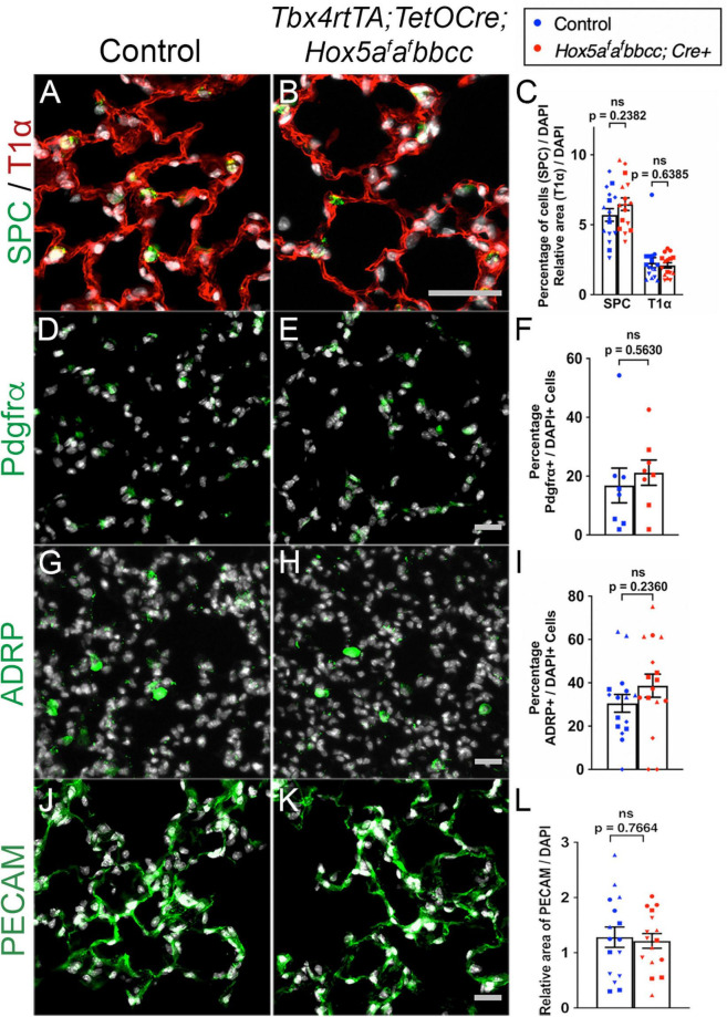
FIGURE 3.
Distal lung cell types are present and appear similar in controls and Hox5 conditional mutants. Lung paraffin sections (A,B,G,H,J,K) and cryosections (D,E) of control and Hox5 adult, conditional mutant mice show similar expression of SPC (green, AECII cells), T1α (red, AECI cells), PDGFRα (green, fibroblasts), ADRP (green, lipofibroblasts) and PECAM (green, endothelial cells); DAPI in gray. Quantification of pixel intensity of T1α (C), and PECAM (L) were normalized to pixel intensity of DAPI per field image. Quantifications of SPC (C), PDGFRα (F) and ADRP (I) cell numbers were normalized to DAPI-positive cell numbers in each panel quantified. Each shape represents an individual animal (ns, not significant). Scale bars: 50 μm. P-values were determined by an unpaired Student’s t-test.

