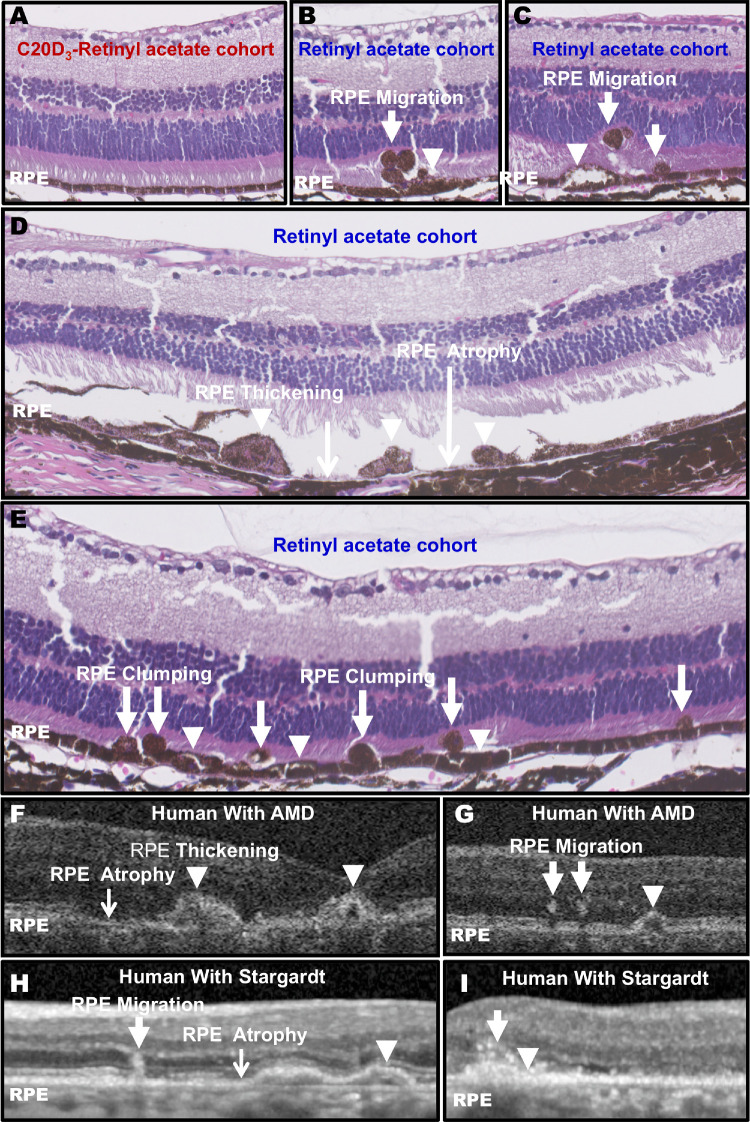
Figure 4.
C20D3-Vitamin A Prevents the Signature RPE Atrophic Changes. (A) Representative hematoxylin and eosin (H&E) cross-section of the C20D3-retinyl acetate cohort at 18 months of age. Histology was performed on n = 12 eyes of 12 mice between 13 and 18 months of age. No RPE pathology was observed in the C20D3-retinyl acetate cohort at any age. (B–E) Representative H&E cross-sections of 18-month-old Abca4−/−/Rdh8−/− animals administered vitamin A as retinyl acetate. Each panel represents a slice from a different eye. RPE migration into the neuroretinal space (arrows), RPE swelling (arrowheads), RPE clumping, or regions of missing or atrophic RPE (black arrows) were observed. Histology was performed on n = 16 eyes of 16 mice between 13 and 18 months of age. All eyes displayed areas of RPE pathology after 13 months of age. (F–I) Representative OTC images of human AMD (F, G) and Stargardt disease (H, I) show similar RPE atrophic changes. Each image is from a different eye.

