Fig. 5.
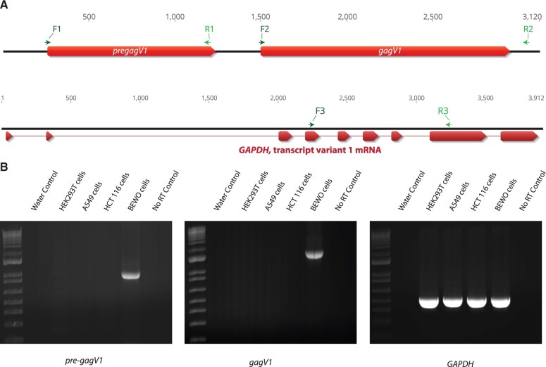
Human pre-gagV1 and gagV1 expression in human cell lines. (A) At the top is a schematic of the genomic region that contains pre-gagV1 and gagV1 ORFs. Locations of the primers used in RT-PCR are indicated above the diagram. At the bottom of the panel is a schematic of the genomic region that contains human GAPDH. Exons of GAPDH are shown in red. Locations of the primers used in RT-PCR are indicated above each diagram. Primers that were designed for different exons of GAPDH were used to confirm the absence of DNA contamination as well as uniform RNA loading. The figure was created using Geneious (Kearse et al. 2012). (B) Agarose gels showing the RT-PCR products of pre-gagV1, gagV1, and GAPDH that were amplified from the indicated human cell lines with the primers shown in the upper panel. A control done without an RT step (only Taq polymerase) was included to confirm the absence of HERV DNA contamination. Gels are representative of at least two independent experiments.

