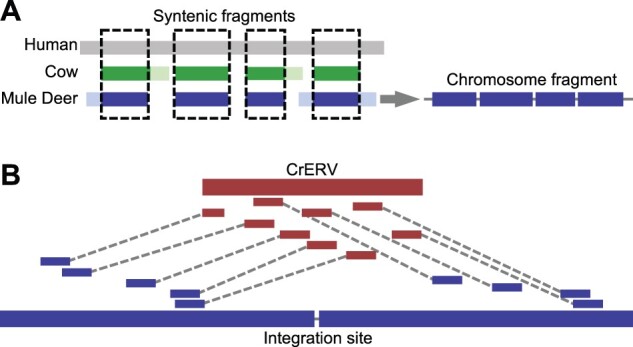
Fig. 1.

Diagram of CrERV reconstruction and RACA. (A) Mule deer chromosome fragment reconstruction using syntenic fragments. Gray, green, and blue boxes correspond to aligned human, cow, and mule deer scaffold respectively. Lighter shades represent regions that can only be aligned between two species. Dashed boxes highlight syntenic fragments where the region is conserved among all three species, which yield a chromosome fragment that orients mule deer scaffolds. (B) Reconstruction of CrERV sequences. CrERV and mule deer scaffolds are shown in bold red and blue boxes, respectively. Long-insert mate pair reads are connected by dotted lines and are colored to indicate whether they derive from the mule deer scaffold or CrERV genome. CrERV genomes were assembled by gathering the broken mate pairs surrounding each CrERV loci as described.
