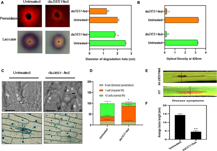
FIGURE 5.
Extracellular enzyme activity and virulence potential of dsDES1-fed strain. (A) The extracellular laccase and peroxidase activity of the in vitro-dsDES1-fed strain on CM plates supplemented with ABTS and Congo red (CR), respectively. The average diameter of halo or discoloration indicator of substrate breakdown was measured in triplicates from 5-day-old plates. (B) Activities of laccases (green) and peroxidases (orange) in culture filtrates of the untreated and dsDES1-fed strain. (C) The upper panel indicates the 40 × -magnified differential interference contrast (DIC) image of onion peel infected with untreated and dsDES1-fed strain. The white arrows indicate the spore, S; appressoria, A; point of penetration, P; invasive hyphal, (IH) growth. The lower panel shows a 10 × -magnified bright field image of Lactophenol blue-stained fungal infection units grown on onion epidermal cells. This is indicative of the cell-to-cell movement of IH and host colonization. (D) Graphical representation of the fraction of infection units that showed differential ease of host-colonization, beyond the primary site of penetration. The potential for cell-to-cell movement of IH was color-coded based on the number of cells they penetrated (0– > 2). At least 50 different infection units were observed for each set, and the experiment was repeated thrice. (E,F) Spot inoculation of detached rice leaves with spores isolated from the in vitro dsDES1-fed strain and untreated wild-type strain. Average length of lesion was measured for each set with approximately 50 leaves (≈ 100 lesions). The error bars represent ± SD, and * and ** denote statistical significance at the P ≤ 0.05 and P ≤ 0.01 levels.

