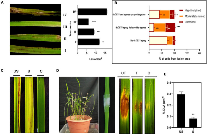
FIGURE 6.
In vivo infection assay of dsDES1-sprayed plants. (A) The left panel shows spray-infected rice leaves that were sandpaper-abraded (to reduce the mechanical barrier) and sprayed with 300 nM dsDES1 (III) 24 h prior, (II) 12 h prior, or (I) 0 h prior (spores and dsDES1 were mixed together before spraying) to fungal inoculation. The unsprayed but fungus-inoculated set (IV) was used as control. The right panel indicates the relative infection severity for each treatment based on lesions/cm2. (B) DAB staining of lesions isolated from treatment sets I, III, and IV, representative of counter-infection host-derived ROS generation assay. The level of ROS generation was color-coded, and the fraction of cells representing each color was categorized. (C) Spray infection assay of dsDES1-sprayed (S) and unsprayed (US) leaves. Leaves that were neither dsRNA-sprayed nor spore-inoculated, were used as control (C). (D) Punch inoculation-mediated infection assay for dsDES1 drop-treated (24 h prior) rice leaves. Leaves that were simply punched and inoculated with Tween 20-Gelatin mixture without spores or dsDES1 were kept as control. (E) Disease severity in dsDES1-sprayed and unsprayed leaves expressed in terms of% diseased leaf Area (DLA). For infection related experiments, observations were performed on at least 50 leaves, and% DLA was assessed using the Image J software. Error bars represent standard deviation (n = 50); where *, **, and *** denote statistical significance (Student’s t-test) at the P ≤ 0.05, P ≤ 0.01, and P ≤ 0.001 levels, respectively.

