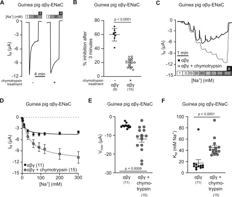
Fig. 7.
Reduced sodium self-inhibition is pivotal to ENaC activity at high extracellular Na+ concentrations. (A) Representative transmembrane current (IM) traces showing sodium self-inhibition (SSI), determined with guinea pig αβγ-ENaC expressing oocytes, with and without prior incubation with chymotrypsin (2 µg/ml in NMDG-ORS for 5 min). Application of amiloride (100 µM) is represented by black bars (a) and [Na+] is represented by white (90 mM) and gray (1 mM) bars. The perfusion speed was 12 ml/min. (B) The percentage of SSI was plotted for guinea pig αβγ-ENaC with and without prior incubation with chymotrypsin (Student’s unpaired t-test). SSI was calculated as (ΔIM peak−ΔIM 3 min)/ΔIM peak×100, where ΔIM peak=IM under 90 mM [Na+] peak −IM under 1 mM [Na+], and ΔIM 3 min=IM after 3 min under 90 mM [Na+]−IM under 1 mM [Na+]. (C) Representative IM traces for guinea pig αβγ-ENaC expressing oocytes with and without prior incubation with chymotrypsin (2 µg/ml for 5 min) across a range of extracellular Na+ concentrations ([Na+]), gray-shaded boxes). (D) The IM from experiments shown in panel (C) were plotted against the extracellular [Na+] and fitted to the Michaelis–Menten equation allowing the estimation of the maximum IM (Vmax) and the [Na+] at which half of Vmax is reached (KM). (E) The Vmax values of guinea pig αβγ-ENaC with and without prior incubation with chymotrypsin (Student’s unpaired t-test with Welch’s correction). (F) The KM values of guinea pig αβγ-ENaC with and without prior incubation with chymotrypsin (Mann–Whitney U test). Numbers in parentheses indicate (n).

