Figure 5.
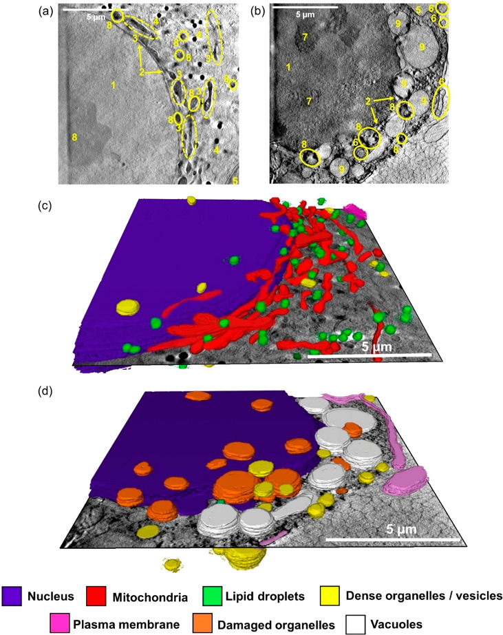
Reconstructed X-ray tomograms and 3D segmented tomograms of two cryopreserved PC3 human prostate cancer cells: untreated control (no drug) under dark conditions (a and c) or exposed to coumarin complex Pt2 after irradiation with blue light (b and d). (a,b) Reconstructed X-ray of PC3 cells exposed to (a) dark conditions for 2 h (Figure S12, Video_T3), or (b) 1× photoIC50 (6.5 μM) Pt2 for 1 h, followed by 1 h blue light (465 nm, 4.8 mW/cm2) irradiation (Figure S21, Video_T16). Distinct cellular features: (1) nucleus; (2) nuclear membrane; (3) mitochondria; (4) lipid droplets; (5) plasma membrane; (6) endosomes/lysosomes; (7) nucleolus; (8) dense organelles; (9) vacuoles. (c-d) 3D segmented tomograms of (a) and (b), respectively (Video_T20 and Video_T21). Images were generated in SuRVoS and visualized in Amira,62 showing subcellular features: nucleus (purple); mitochondria (red); lipid droplets (green); dense organelles/vesicles (yellow); plasma membrane (magenta), damaged (unidentifiable) organelles (orange), and vacuoles (white). Significant cellular damage can be observed in (d) compared to the untreated controls (c) including blebbing of the plasma membrane, damage to organelles in the cytoplasm and nuclear membrane, presence of cytoplasmic vacuoles, and reduced number of lipid droplets.

