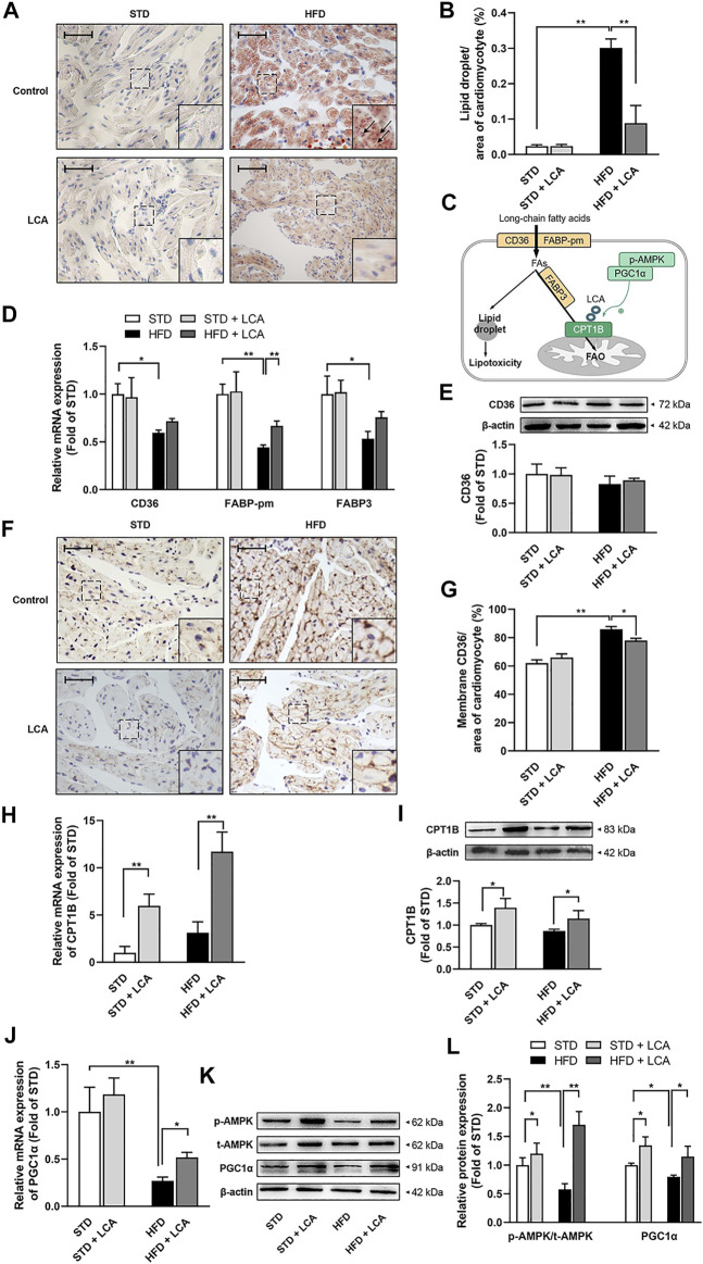
FIGURE 3.
LCA enhances FAO, thereby inhibits cardiac lipotoxicity by alleviating atrial steatosis in obese mice. (A) Representative sections of lipid accumulation (Arrow) using oil red O staining. (B) Quantitative analysis of lipid accumulation by ImageJ. (C) Schematic diagram illustrating cardiac lipid metabolism. (D) Quantitative analysis of the transcription of lipid uptake and transportation-related genes (CD36, FABP-pm, FABP3) by RT-PCR. (E) Representative image and quantitative analysis of CD36 by Western blot. (F) Representative images of membrane translocation of CD36 using immunohistochemistry. (G) Quantitative analysis of sarcolemma CD36 contents by ImageJ. (H) Quantitative analysis of the transcription of CPT1B using RT-qPCR. (I) Representative Image and quantitative analysis of CPT1B by Western blot. (J) Quantitative analysis of the transcription of PGC1α using RT-qPCR. (K,L) Representative images and quantitative analysis of FAO-related regulators (AMPK and PGC1α) by Western blot. Scale bar: 50 μm n = 4 per group. One-way ANOVA with Bonferroni post-hoc test was used to compare data among groups. Data are expressed as mean ± SEM. * p < 0.05, ** p < 0.01. STD, standard diet; HFD, high-fat diet; LCA, l-carnitine; FAs, fatty acids; FAO, fatty acids oxidation; AMPK, AMP-activated protein kinase; CD36, FAT; CPT1B, carnitine palmitoyltransferase-1B; PGC1α, peroxisome proliferator-activated receptor γ coactivator1α; FABP-pm, plasma membrane fatty acid-binding protein; FABP3, fatty acid binding protein 3; p-, phoso-.

