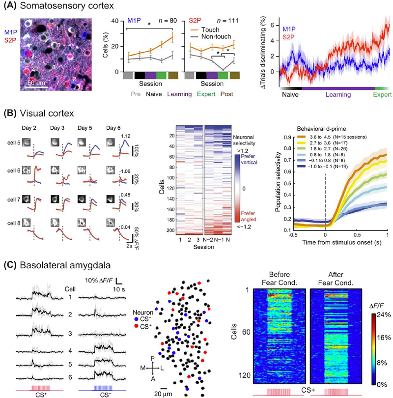
Figure 3.
Learning-related changes in neuronal population activity measured with chronic cellular imaging. A. Projection neurons in primary somatosensory cortex (S1) show distinct learning-related activity during performance of a tactile discrimination task. Left: Identification of M1-projecting (M1P) and S2-projecting (S2P) neurons via retrograde tracers. Middle: The fraction of neurons classified as touch or non-touch as a function of Naive, Learning, and Expert behavioral phases. Right: The change in discrimination accuracy of M1P and S2P neurons for Go and NoGo tactile stimuli (P100 vs. P1200 textures) through learning. Adapted from (Chen et al. 2015a). B. Neurons in primary visual cortex (V1) show diverse learning-related activity during performance of a visual discrimination task. Left: Example calcium signals from four neurons across four imaging sessions in response to a vertical, rewarded stimulus (blue) or an angled, non-rewarded stimulus (red). Middle: Selectivity of neurons across the first three and last three training sessions. Right: Neuronal population selectivity as a function of learning. Each curve depicts the time course of selectivity at a range of behavioral d’. Adapted from (Poort et al. 2015). C. Neurons in basolateral amygdala change selectivity with auditory fear conditioning. Calcium signals of cells responsive to two different auditory tones (CS+ or CS−) before pairing the conditioned stimulus (CS+) with a foot shock using a fear conditioning paradigm. Right, Cell population responses in an example mouse to the CS+ tone before and after fear conditioning. A, anterior; L, lateral; M, medial; P, posterior. Adapted from (Grewe et al. 2017).

