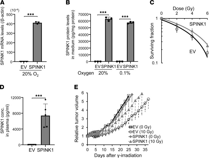
Figure 4. SPINK1 accelerates tumor growth after radiotherapy.
(A–C) DU145/EV and DU145/SPINK1 cells were cultured under the indicated oxygen conditions for 48 hours and subjected to qPCR (A) and the ELISA assay (B) or treated with the indicated dose of γ-ray irradiation for the clonogenic survival assay (C). (D and E) DU145/EV or SPINK1 xenografts were locally irradiated at a dose of 0 (solid lines) or 10 (dotted lines) Gy. When the volumes of the xenografts reached the same sizes as those on day 0, plasma SPINK1 levels were quantified by ELISA assays (D). Tumor growth was analyzed after the treatment (E). Data are represented as mean ± SD (n = 3 in A and B, n = 6 in C, n = 5 in D, and n = 9–10 in E). Two-tailed Student’s t test. ***P < 0.001. SPINK1, serine peptidase inhibitor Kazal type 1; EV, empty vector.

