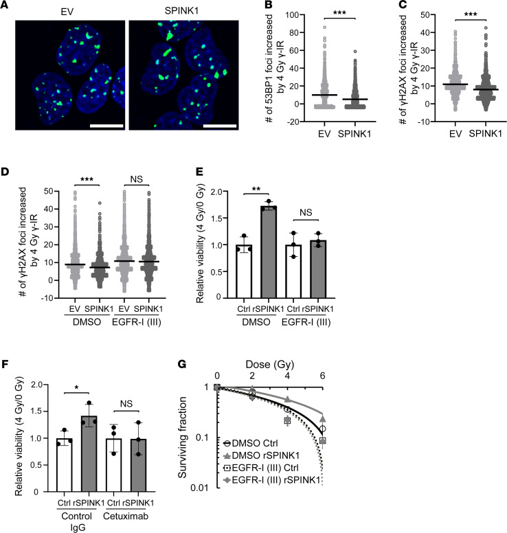
Figure 5. SPINK1 decreases radiation-induced DNA damage and enhances radioresistance of cancer cells in a EGFR-dependent manner.
(A–D) Four days after being transfected with either pcDNA4/SPINK1 or its EV, DU145/EGFP-53BP1-M (A and B) and DU145 (C and D) cells were irradiated with 0 or 4 Gy of γ-rays in the presence or absence of EGFR-I III (D), and the DNA DSBs detected as EGFP-53BP1 foci (A and B) or as γH2AX foci (C and D) were analyzed 2 hours (A and B) or 15 minutes (C and D) after the radiation. (A) Immunocytochemical analysis. Green, EGFP-53BP1 foci; blue, counter staining using Hoechst 33342. Scale bar: 10 μm. (B–D) The number of foci increased by 4 Gy γ-IR was calculated by subtracting the number of foci at 0 Gy from that at 4 Gy under each condition and represented as dot plots with mean ± SD. (E and F) DU145 cells were irradiated with 0 or 4 Gy of γ-ray in the presence or absence of 100 ng/mL rSPINK1 in combination with DMSO or 0.5 μM EGFR-I III (E), or with control IgG or 10 μg/mL cetuximab (F), and subjected to colorimetric cell viability assays. (G) The same experiment as in Figure 3B was conducted in the presence or absence of EGFR-I III. Data are represented as mean (n > 1000 in B–D) and mean ± SD (n = 3 in E and F, and n = 6 in G). Two-tailed Student’s t test. *P < 0.05, **P < 0.01, ***P < 0.001. SPINK1, serine peptidase inhibitor Kazal type 1; EV, empty vector. EV, empty vector; EGFR-I III, EGFR Inhibitor III; DSBs, double-strand breaks.

