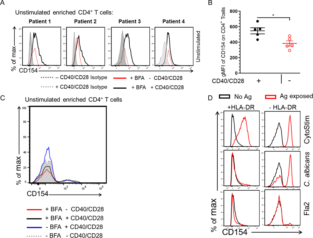
Figure 3. Identifying factors that contribute to increase CD154 background on unstimulated cells.
(A) Unstimulated PBMCs were incubated for 7hr in the presence or absence of anti-CD40/anti-CD28 with BFA. Cells were enriched and stained for CD154 expression. Histogram plots of 4 donors showing CD154 expression among viable CD4+ T cells. (B) Scatter plot represents gMFI of CD154 expression in CD14−CD20−CD8−CD4+ T cells incubated with (black circle) or without anti-CD40/anti-CD28 (red circle) in the presence of BFA. Bars represent the means ± SEM of five independent experiments using different Crohn’s patients (n = 5); Mann-Whitney U comparison. *p<0.05. (C) Histogram of CD154 expression of a representative Crohn’s unstimulated CD4+ T cells incubated for 7hr with or without anti-CD40/anti-CD28 in the presence or absence of BFA. (D) Antigen exposed PBMCs from one active Crohn’s patient cultured in the presence of anti–HLA-DP-DR-DQ.

