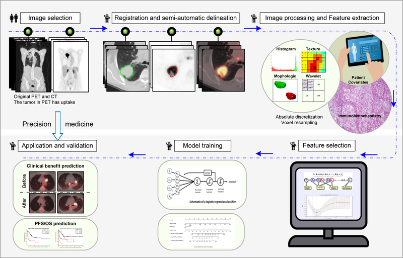
Fig 1. Radiomics Workflow.
The workflow includes image selection (only images with slice thickness≤5 mm no artifacts, and the tumor in PET images has FDG uptake were included), registration and automatic delineation, imaging preprocessing and feature extraction, feature selection, model training and model validation.

