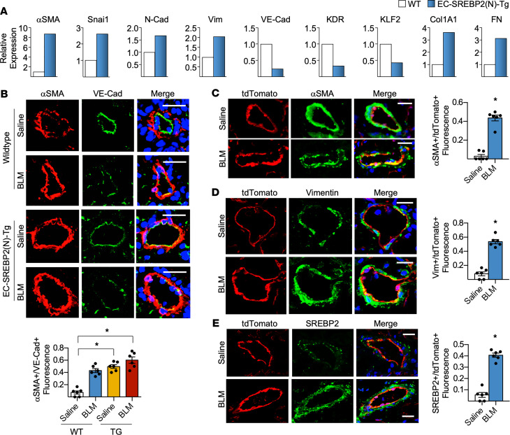
Figure 6. BLM-induced SREBP2 promotes EC phenotypic switch and partial EndoMT in mice.
(A) Lung microvascular ECs were isolated from age-matched EC-SREBP2(N)-Tg mice and compared with their WT littermates (pooled from n = 5 per group). The level of indicated mRNA was measured using qPCR. (B–E) Age-matched EC-SREBP2(N)-Tg and WT littermates, or EC-specific tdTomato-expressing mice driven with tamoxifen inducible VE-Cad–Cre (EC-tdTomato mice) were administered BLM (8 units/injection) on days 0, 4, 7, 10, and 14 through i.p. injection. Twenty-eight days after BLM administration, lungs were harvested. (B) Frozen lung sections were immunostained for the EC marker VE-Cad (green) and the mesenchymal marker αSMA (red). Nuclei were counter stained with DAPI (blue). Scale bar: 20 μm. Quantitative analysis showing the number of αSMA+ cells compared with total VE-Cad+ cells in the intima is graphed below the representative images. (C–E) Lineage-tracing experiments were performed with EC-specific expression of tdTomato (red), counterstained with αSMA (green) (C), vimentin (green) (D), or SREBP2 (green) (E). Nuclei are labeled with DAPI (blue). Scale bar: 20 μm. Quantitative analysis showing αSMA+, Vim+, or SREBP2+ cells are compared with total tdTomato+ cells in the intima, which is graphed on the right of the representative images. Data in B were analyzed by 2-way ANOVA with Kruskal-Wallis post hoc; data are represented as mean ± SEM from n = 6 mice per group. Data in C–E were analyzed by 2-tailed Student’s t test; data are represented as mean ± SEM from n = 6 mice per group. *P < 0.05 between the indicated groups. Col1A1, collagen 1 type 1; FN1, fibronectin 1; KDR, kinase insert domain receptor; KLF2, Krüppel-like factor 2; N-Cad, neural cadherin; Snai1, snail family transcriptional repressor 1; VE-Cad, vascular endothelial cadherin; Vim, vimentin; Wnt, wingless integration site).

