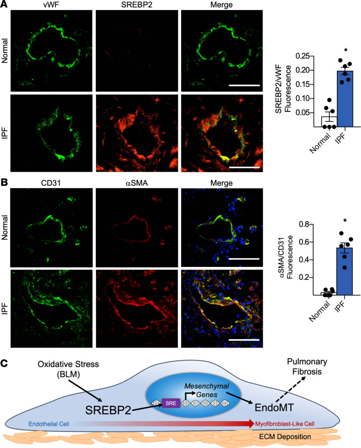
Figure 8. SREBP2 and mesenchymal markers are induced in human lung endothelium with idiopathic pulmonary fibrosis (IPF).
(A and B) Human lung tissue samples from patients with IPF were compared with normal lung transplant controls (n = 6 per group). Immunostaining was performed with the endothelial cell marker vWF (green) and SREBP2 (red) (A), or the endothelial cell marker CD31 (green) and mesenchymal marker αSMA (red) (B). In all images, nuclei were counter stained with DAPI (blue). Scale bar: 20 μm. Data in A and B were analyzed by 2-tailed Student’s t test; data are represented as mean ± SEM from n = 6 patient samples per group. *P < 0.05 between the indicated groups. BLM, bleomycin; CD31, PECAM-1; EndoMT, endothelial-to-mesenchymal transition; vWF, von Willebrand factor. (C) The involvement of BLM/SREBP2/EndoMT axis in the lung ECs during the onset of PF.

